Seeing The Spread: Researching How To Make Operating Rooms Safer With Help of FLIR GF343

Ensuring the safety of surgical staff includes having high air quality in the operating room. Proper air regulation decreases the spread of air-borne infections—including COVID-19—between patients and staff. Analyzing how those infections spread is of key interest to researchers at the University of Dublin, in Ireland, who are investigating how the use of carbon dioxide (CO2) in minimally invasive surgery might contribute to infection rates among doctors and surgery staff. In order to study the effect of CO2 in surgery, the researchers brought in the FLIR GF343, an optical gas imaging (OGI) camera capable of visualizing CO2. Before we dig into how or why they used this camera, though, it is important to note that the FLIR GF343 is not intended, marketed, or sold for use in the medical field or surgery. In this case, researchers used the camera to better understand the surgery setting.

FLIR GF343 Camera
COVID-19 Fears Among Surgery Staff
Evidence suggests that 30% of coronavirus infections among health care staff resulted from exposure to sick patients who spread the virus through aerosolized particles from sneezing, coughing, and speaking. Efforts to protect surgery staff from exposure currently include additional layers of protective, sterile clothing (PPE) as well as methods such as positive pressure ventilation and continuous air exchange in operating rooms. This air quality can be compromised, though, by the amount of equipment, number of personnel, and level of emissions. Among these factors, there was concern that cautery smoke, aerosolized gases, liquids, chemicals, and pathogen particles could spread infections in the OR. But with the cancellation of non-emergencies surgeries during heavy COVID-19 quarantining, professionals had the opportunity to study new safety measures to lower infection rates. This is when doctors became curious about the amount of medical gas leaking during minimally invasive surgery (MIS) and its role in spreading infections.
CO2’s Role in Surgery
MIS, or keyhole surgery, is a procedure for accessing the interior of the body through a small incision. MIS frequently involves the use of medical grade CO2 to enlarge and stabilize body cavities for better visibility and instrument maneuverability during procedures. CO2 works well because the gas is nonflammable, inexpensive, and has higher blood solubility than air. The only issue though is that escaping gas, cautery smoke, and aerosolized cells spreading via gas leaks are an inherent risk when using gas in a surgery setting.
Despite screening patients prior to their operations, evidence early in the pandemic suggested that COVID-19 was still present in blood and stool in colorectal surgeries. Combine this with concerns about escaping gas and you have a very real fear that the gas leaks were carrying infectious particles to surgery staff.
Investigating the Potential Hazards
The fear that CO2 gas leaks were contributing to infections piqued the interest of Professor Ronan Cahill, MD and Professor of Surgery at Mater Misericordiae Hospital and University College of Dublin. “The surgeon in me presumed the leaks were insubstantial; the academic in me wanted to quantify and demonstrate the truth,” says Cahill. “I consulted with Dr. Kevin Nolan, who lectures and teaches in the School of Materials and Mechanical Engineering at UCD.”
Nolan has extensive experience in Schlieren imaging, an imaging technique popular in aeronautic testing that visualizes local changes in the refractive index of air. “I had performed Schlieren testing for effluvium (gas leakage) coming from an insufflated body cavity,” Nolan explains. “Part of the process involved class four lasers on a surgical simulation models to illuminate the particles and capturing them with a super slow motion Phantom camera. But that is a complex setup in an OR.” While particles became visible, the use of lasers rendered this method impractical and dangerous in a live human setting.
Nolan and Cahill determined they had to find another way to visualize the medical gas. Both had coincidently seen a documentary on climate change called “Racing Extinction” by Louie Psihoyo, which features thermal imaging with a specialized filter that made everyday carbon dioxide emissions visible. Cahill contacted the director to learn more about the technology they used and to determine whether it could aid their research.
A Practical Solution
Now that Cahill and Nolan had a new lead to follow, the two went to quick work obtaining grants and funding for their research and the purchase of a FLIR GF343 gas imaging camera. The benefits of the camera were immediately apparent: the GF343 was less obtrusive and far easier to setup than the previous Schlieren approach. Cahill’s main objective was to observe leakage while access devices called “trocars” and surgical valves were in place. Surgeons use trocars and valves in colorectal and other abdominal surgeries for inserting, guiding, and retracting specialized instruments. The GF343 made the leaks clear as day; the camera showed normally-invisible CO2 pluming out of the instruments and flowing over a wide area, increasing as the fit of the valves naturally loosened over the course of a surgery.

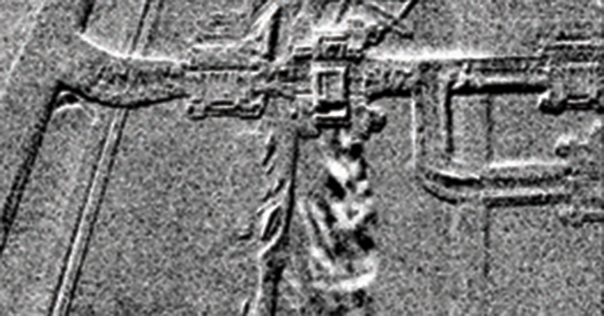
Footage from Cahill and Nolan showing CO2 leaking out of the instrument and leaving a trail across the dummy arm.
Cahill’s original goal was to visualize the amount of gas leakage happening during surgery but these results exceeded even his expectations. The research confirmed that surgery teams are exposed to significantly more leakage and particles than previously estimated. The much larger, overarching goal, though, was to raise awareness for additional efforts needed to protect patients and medical professionals from the spread of viruses in breathing zones. Cahill claims that surgeons who spend limited time in a surgery environment were generally not too worried, nurses and other staff who visit multiple procedures a day were glad to know their concerns were being taken seriously.
The research has been performed across different surgical teams and specialties at the Mater Misericordiae University Hospital in Dublin, Ireland as well as IRCAD-EITS, Strasbourg, France as part of a consortium award by EU Horizon 2020 called “Protecting Operating Rooms Staff against Aerosolised Viruses (PORSAV)”.
Related Articles
-
 Application Story
FLIR Optical Gas Imaging Camera helps AGL Energy reduce hydrogen leak inspection time in power station by 50%
Read the story -
 Download
Optical Gas Imaging: The Professional Guide
Read the Story -
 Fundamentals
How Sensitive are FLIR Gas Detection Cameras?
Learn more